
Fracture Patterns and Goal of Treatment
A well-aligned and stable supporting skeleton is required for the stability of small articulations of hand, for the balance between its extrinsic and intrinsic motors, and for the complexity of its tendon systems. However, on the return of function of these soft-tissue structures on the skeletal union, the end-result of the hand’s skeletal injuries can be judged better.
One must try to obtain the patient’s cooperation for the assessment of the pre-operative status of the tendon, motor, sensory, and vascular, and all that should be documented carefully. The goals for the treatment of meta-carpal and phalangeal fractures are the same no matter what method is used. The goals are:
- Restoration of articular anatomy
- Stabilization of fractures.
- Eradication of angular or rotational deformity.
- Surgically acceptable wounds.
- Expeditious mobilization.
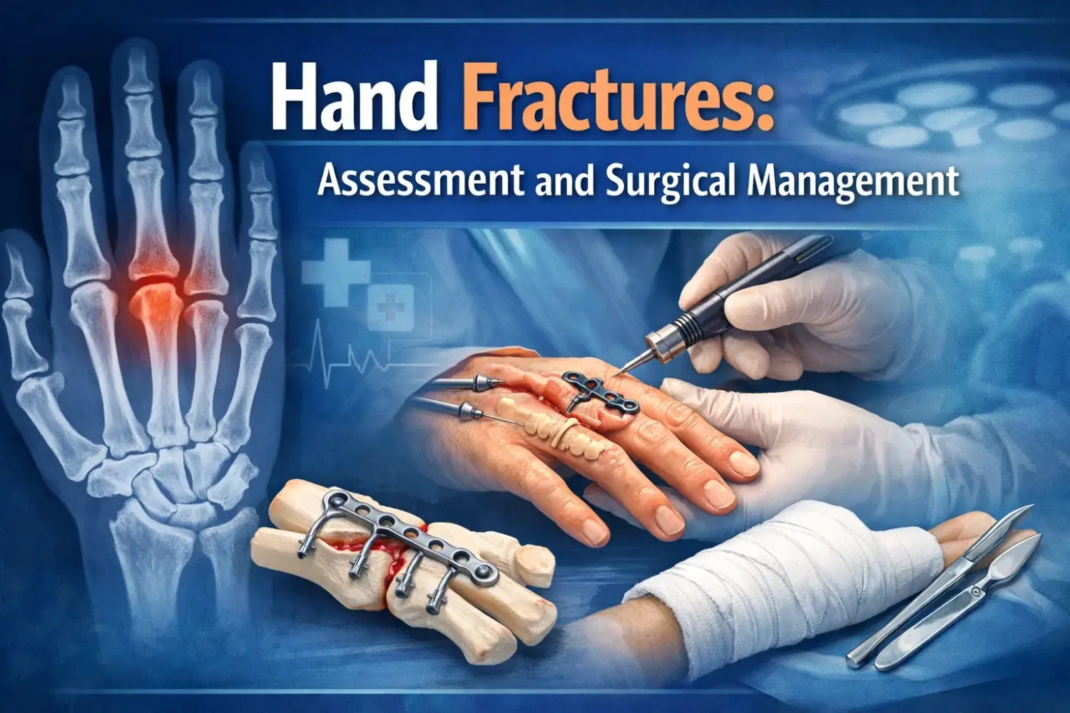
Though non-operative means can be used effectively to treat fractures of the tubular skeleton in the hand. But Some fractures, due to their situation, nature, or associations, need stable skeletal fixation. Such fractures are multi-fragmented fractures, multiple metacarpals, spiral metacarpal associated with soft tissue injury, severely displaced fractures, short oblique fracture; fractures of various sites as proximal phalanx, sub-condylar and palmar based middle phalanx; displaced articular fractures: Bennett’s and Rolando’s uni-condylar and bi-condylar fractures; fractures associated with specific injuries as complete or incomplete amputations and fracture-dislocations.
In the above-mentioned fractures, the location, and pattern of the fracture, its response to a motion, and associated injuries determine the stability. Locking plates for hand fractures (titanium) are used for different hand fractures. These plates allow free choice for screw placement which is a major advantage in case of complex fractures.
Surgical anatomy
The breadth of the hand is formed by the four metacarpals and a rigid central pillar which is extending through the index and middle metacarpals. The distal transverse arch of the hand is located along the deep metacarpal ligaments interlinking the metacarpal heads. The mobile units are the thumb and the ring and little finger metacarpals. The metacarpal shaft distally extends with a gentle dorsal convexity. The concave palmar cortex is denser in comparison to the dorsal cortex. This suggests a compression side on the palmar aspect, generally with tensile stresses which present dorsally.
Contrary to the metacarpals, the phalanges are wrapped by the gliding surfaces of the overlying intrinsic and extrinsic tendons. The proximal and the structure of middle phalanges are divided into the base, shaft, neck, and head (condyles). The configuration of the distal articular anatomy of the proximal interphalangeal joint is condylar which seems like a grooved trochlea. The width of the palmar aspect is about twice of the dorsal margin. Compressive forces give rise to dynamic stability within a joint that is increased by pinch and grip, whereas passive stability derives from collateral ligament tension which is increased by joint flexion.
The metacarpal head shows a curve of increasing diameter when it is viewed in the sagittal plane but it is in pear shape if viewed in the coronal plane. The joint remains stable in flexion and lax in extension due to the peculiar origin of the collateral ligaments on that oddly shaped metacarpal head. The carpometacarpal joint of the thumb is a reciprocally biconcave saddle joint. The relationship of the reciprocal concave and convex arcs of the bases (almost at 90° to each other) of its metacarpal and trapezium allows the thumb’s free movement at an extensive degree.